Perio-Implant Journals

跟牙周病治療與植牙相關的文獻

My Photo
Name:
Location: 台南市, Taiwan

A tour-crazy periodontist around the World. - I like to drive VOVLE cars and put sweden-made dental implants (Nobelcare & Astra) and live with IKEA furnishes .

Monday, August 13, 2007

Triple Immediate Therapy (ridge expansion, soft tissue augmentation, and provisional restoration) of Maxillary Anterior Single Implant

Journal of Periodontology
2007, Vol. 78, No. 7, Pages 1348-1353
(doi:10.1902/jop.2007.060487)

Yu-Lin Lai,* ­I-Chiang Chou,* ­Yu-Cheng Liaw,* ­Hen-Li Chen,* ­Yi-Chun Lin,* and ­Shyh-Yuan Lee*­

*Department of Dentistry, Taipei Veterans General Hospital, Taipei, Taiwan.

†School of Dentistry, National Yang-Ming University, Taipei, Taiwan.

‡Department of Dentistry, Taipei City Hospital, Taipei, Taiwan.

Correspondence: Dr. Shyh-Yuan Lee, School of Dentistry, National Yang-Ming University, Taipei 112, Taiwan. Fax: 886-2-28264053; e-mail: sylee@ym.edu.tw.

Background: Immediate restoration during implant placement in the premaxillary area can achieve a favorable esthetic result. However, the treatment always poses a great challenge to clinicians, especially for patients with preexisting soft and hard tissue deficiencies. The aim of this clinical report was to present a protocol for a multidisciplinary approach to provide an immediate implant restoration for patients with ridge deficiency.

Methods: A 21-year-old female presented with a mesial shift of the maxillary right canine due to loss of a lateral incisor at an early age. The treatment protocol included orthodontic space redistribution to open a canine space for implant placement. Subsequently, ridge expansion and implant placement combined with connective tissue grafting for the reconstruction of the soft tissue profile were performed, and an immediate provisional prosthesis was delivered simultaneously. A definitive University of California at Los Angeles–style ceramometal crown was completed 6 months later with periodical clinical maintenance.

Results: The 18-month postoperative follow-up revealed that the implant was stable, and the buccal depression of the surgical area was reconstructed. Papillae were augmented, and a harmonious soft tissue margin was achieved in the esthetic zone. Radiographs demonstrated a normal vertical osseous height and excellent osseointegration of the implant.

Conclusions: The combination technique of ridge expansion and soft tissue augmentation can be applied to immediate implant restoration cases without the need for complicated bone grafting surgeries in atrophic ridges. This triple immediate strategy enabled us to reduce the number of surgeries, which simplified the overall procedure without compromising the esthetic results.

Labels:

Sunday, February 25, 2007

Interventions for replacing missing teeth: bone augmentation techniques for dental implant treatment

Esposito M, Grusovin MG, Worthington HV, Coulthard P

The Cochrane Database of Systematic Reviews 2007 Issue 1
Copyright © 2007 The Cochrane Collaboration. Published by John Wiley and Sons, Ltd.

Date of last Subtantial Update: February 21. 2006

Authors' conclusions

Major bone grafting procedures of extremely resorbed mandibles may not be justified. Bone substitutes (Bio-Oss or Cerasorb) may replace autogenous bone for sinus lift procedures of extremely atrophic sinuses. Both guided bone regeneration (GBR) procedures and distraction osteogenesis can augment bone vertically, but it is unclear which is the most efficient technique. It is unclear whether augmentation procedures at immediate single implants placed in fresh extraction sockets are needed, and which is the most effective augmentation procedure, however, sites treated with barrier + Bio-Oss showed a higher position of the gingival margin, when compared to sites treated with barriers alone. Non-resorbable barriers at fenestrated implants regenerated more bone than no barriers, however it remains unclear whether such bone is of benefit to the patient. It is unclear which is the most effective technique for augmenting bone around fenestrated implants. Bone morphogenetic proteins may enhance bone formation around implants grafted with Bio-Oss. The use of particulated autogenous bone from intraoral locations, also taken with dedicated aspirators, might be associated with an increased risk of infective complications. These findings are based on few trials including few patients, having sometimes short follow up, and being often judged to be at high risk of bias.

Labels: ,

Augmentation procedures for the rehabilitation of deficient edentulous ridge with oral implant

Matteo Chiapasco, Marco Zaniboni, Maurizio Boisco,
Clin. Oral Impl. Res. 17 (Suppl. 2), 2006; 136–159

Unit of Oral Surgery, Department of Medicine, Surgery, and Dentistry, San Paolo Hospital, University of Milan, Milan, Italy

Results: Success rates of surgical procedures ranged from 60% to 100% for GBR, from 92%
to 100% for onlay bone grafts, from 98% to 100% for ridge expansion techniques, from
96,7% to 100% for DO, and was 87.5% for revascularized flaps, whereas survival rates of
implants ranged from 92% to 100% for GBR, from 60% to 100% for onlay bone grafts, from
91% to 97.3% for RE, from 90.4% to 100% for DO, and, finally, was 88.2% for
revascularized flaps.
Conclusion: On the basis of available data it was shown that it was difficult to demonstrate
that a particular surgical procedure offered better outcome as compared to another. The
main limit encountered in this review has been the overall poor methodological quality of
the published articles. Therefore larger well-designed long term trials are needed.

Labels: ,

Effects of zinc deficiency on oral and periodontal diseases in rats

Department of Periodontology, Faculty of Dentistry, and
2Department of Histology and Embryology, Medical School, Atatürk
University, Erzurum, Turkey

Dr Cankat Kara, Atatürk Üniversitesi Diş Hekimliği Fakültesi,
Periodontoloji Anabilim Dalı 25240, Erzurum, Turkey
Tel: +90 442231 1902
Fax: +90 442212 2380
e-mail: mcankat@hotmail.com

Conclusion: The findings indicated that oral health was better in
group II rats (those fed with a zinc-containing diet) than in group I
(zinc-deficient) rats. Hyperkeratinization was more prominent in
zinc-deficient rats. We suggest that zinc deficiency is a potential
risk factor for oral and periodontal diseases.

Labels:

Saturday, February 17, 2007

Factors Affecting Late Implant Bone Loss: A Retrospective Analysis

Factors Affecting Late Implant Bone Loss: A Retrospective Analysis

Dyeus M. Chung, DDS, MS / Tae-Ju Oh, DDS, MS / Jungwha Lee, MS / Carl E. Misch, DDS, MDS / Hom-Lay Wang, DDS, MSD


Purpose: Prevention of late implant bone loss is a critical component in long-term success of implants. The aim of the present study was to evaluate factors affecting late implant bone loss.
Materials and Methods: Three hundred thirty-nine endosseous root-form dental implants placed between April 1981 and April 2002 in 69 patients were analyzed. The implants were categorized based on the following factors: (1) surface characteristics (smooth versus rough), (2) length (short [< 10 mm] versus long [≡ 10 mm]), width (narrow [< 3.75 mm], regular [3.75 to 4.0 mm], or wide [> 4.0 mm]), (3) the amount of keratinized mucosa (< or ≡ 2 mm), (4) location (anterior versus posterior; maxilla versus mandible), (5) type of prosthesis (fixed versus removable), and (6) type of opposing dentition. The effects of these factors on clinical parameters, especially average annual bone loss (ABL), were evaluated clinically and radiographically by a blinded examiner. The parameters evaluated were modified Plaque Index, Gingival Index, modified Bleeding Index, probing depth, and ABL.
Results: Shorter implants, wider implants, implants supporting fixed prostheses, and implants in smokers were found to be associated with greater ABL (P < .05). The random intercept mixed effects model showed that implant length was the most critical factor for maintenance of ABL.
Conclusions: Shorter implants, wider implants, implants supporting fixed prostheses, and implants in smokers were associated with greater ABL. Implant length was the most significant factor in the maintenance of dental implants. Randomized controlled clinical trials are needed to confirm the results obtained from this retrospective clinical study. (Case Series) (More than 50 references.) Int J Oral Maxillofac Implants 2007;22:117每126

Key words: implant maintenance, implant surfaces, late implant bone loss, peri-implantitis

Labels:

Saturday, January 20, 2007

Vertical ridge augmentation with guided bone regeneration in association with dental implants: an experimental study in dogs

Authors: Simion, Massimo1; Dahlin, Christer2; Rocchietta, Isabella1; Stavropoulos, Andreas3; Sanchez, Raquel4; Karring, Thorkild3

Source: Clinical Oral Implants Research, Volume 18, Number 1, February 2007, pp. 86-94(9)

Publisher: Blackwell Publishing

Abstract:
Aim:

To evaluate the effect of using guided bone regeneration (GBR) with a titanium-reinforced e-PTFE membrane in alveolar bone defects with titanium implants. Material and methods:

Following extraction of three mandibular premolars and a molar on both sides of the jaw in three dogs, alveolar bone defects (depth: 5-7 mm) were produced. After 4 months, three implants were inserted into each defect to a depth of approximately 4 mm, so that their coronal portion was protruding about 5 mm. Four sides in the dogs were assigned to a test group and the remaining two sides to a control group. The 12 implants in the test group were covered with a reinforced e-PTFE membrane. The space under the membrane was filled with peripheral venous blood from the animal, and the flaps were sutured over the membrane. The six control implants received no membrane before the suturing of the flaps to complete wound closure. The animals were sacrificed after 6 months, and non-decalcified histological specimens of the implants and surrounding tissues were prepared. Results:

Histologic and histomorphometric analyses revealed a significantly (Mann-Whitney test; P=0.08) larger amount of bone fill in the test group (mean=57.42%) than in the controls (mean=11.65%), and clinical evaluation of one test site showed that the implants were completely covered with tissue resembling bone. In most of the specimens, bone had grown in height close to, or in direct contact with the membrane. However, the new bone generally was not in direct contact with the implants. Regularly, a zone of dense connective tissue was interposed between the implants and the newly formed bone.

Conclusion:

The formation of even considerable amounts of bone following vertical ridge augmentation with GBR and implants was not accompanied by predictable osseointegration of the implants.

Fully vs. partially rough implants in maxillary sinus floor augmentation: a randomized-controlled clinical trial

Authors: Stavropoulos, Andreas1; Karring, Thorkild1; Kostopoulos, Lambros2

Source: Clinical Oral Implants Research, Volume 18, Number 1, February 2007, pp. 95-102(8)

Publisher: Blackwell Publishing

Abstract:
Objectives:

To compare implants with a rough surface in their whole length (FR) with implants having a 2 mm coronal machined portion (PR) when used in association with a sinus-lift procedure. Material and methods:

Twenty-six patients with 2 mm≤x≤9 mm residual alveolar crest were prosthetically restored with implants after a staged sinus-lift procedure using osteotomes. In 13 randomly chosen patients, no more than one FR implant was placed (test group), while the rest were PR implants. The other 13 patients received only PR implants (control group). For comparisons, only one implant from each patient was used, i.e., from the test group only the 13 FR implants were used, while from the control group, one PR implant was randomly chosen. The presence/absence of plaque, BOP, PPD and REC were registered at the day of delivery of the restorations and after 1 year. Residual alveolar crest height and marginal bone levels around the implants were evaluated on standardized periapical radiographs taken at various stages. Results:

Four FR and two PR implants were lost, and the cumulative survival rate was 82.9% (six lost out of 35). There were no significant differences between the two groups. Implant type, residual alveolar crest height, time of osseointegration, time of implant loading and smoking did not seem to influence implant survival. Conclusions:

FR and PR implants placed in augmented sinuses did not differ in their clinical performance.

Wednesday, April 19, 2006

Biological Width for Dental Implants

J Periodontol 1997 Feb;68(2):186-98
Biologic width around titanium implants.
A histometric analysis of the implanto-gingival junction around unloaded and loaded nonsubmerged implants in the canine mandible.
Cochran DL, Hermann JS, Schenk RK, Higginbottom FL, Buser D
Department of Periodontics, Dental School, University of Texas Health Science Center at San Antonio, USA.
The use of endosseous dental implants as transmucosal devices necessitates the successful integration of three different tissues: bone, connective tissue, and epithelium. So far, studies have predominantly focused on hard tissue integration. Much less is known about soft tissues. This study examined the dimensions of the implantogingival junction in relation to clinically healthy unloaded and loaded nonsubmerged implants. In total, 69 titanium plasma-sprayed (TPS) and sandblasted acid-etched (SLA) implants were placed in an alternating fashion in six foxhounds and allowed to heal for 3 months. Two dogs were sacrificed after the initial healing period. The remaining four dogs had crowns fabricated that were allowed to function for up to 12 months. These animals were sacrificed after 3 and 12 months of loading. Histometric analysis of undecalcified histologic sections included the evaluation of the sulcus depth (SD), the dimensions of the junctional epithelium (JE), and the connective tissue contact (CTC). Mean values in the 3 month unloaded group were 0.49 mm for SD, 1.16 mm for JE, and 1.36 mm for CTC. These dimensions were 0.50 mm for SD, 1.44 mm for JE, and 1.01 mm for CTC for the 3 month loaded group. After 12 months of loading, these values were 0.16 mm for SD, 1.88 mm for JE, and 1.05 mm for CTC. The sum of these measurements was similar for the different time points and similar to the same dimensions around teeth. TPS and SLA surfaces had no influence on the evaluated parameters (P > 0.05). The data suggest that a biologic width exists around unloaded and loaded nonsubmerged one-part titanium implants and that this is a physiologically formed and stable dimension as is found around teeth.
PMID: 9058338, UI: 97211340

J Biomed Mater Res 1998 Apr;40(1):1-11
Bone response to unloaded and loaded titanium implants with a sandblasted and acid-etched surface: a histometric study in the canine mandible.
Cochran DL, Schenk RK, Lussi A, Higginbottom FL, Buser D
Department of Periodontics, University of Texas Health Science Center at San Antonio 78284-7894, USA. cochran@uthscsa.edu
Many dental clinical implant studies have focused on the success of endosseous implants with a variety of surface characteristics. Most of the surface alterations have been aimed at achieving greater bone-to-implant contact as determined histometrically at the light microscopic level. A previous investigation in non-oral bone under short-term healing periods (3 and 6 weeks) indicated that a sandblasted and acid-etched titanium (SLA) implant had a greater bone-to-implant contact than did a comparably-shaped implant with a titanium plasma-sprayed (TPS) surface. In this canine mandible study, nonsubmerged implants with a SLA surface were compared to TPS-coated implants under loaded and nonloaded conditions for up to 15 months. Six foxhound dogs had 69 implants placed in an alternating pattern with six implants placed bilaterally in each dog. Gold crowns that mimicked the natural occlusion were fabricated for four dogs. Histometric analysis of bone contact with the implants was made for two dogs after 3 months of healing (unloaded group), 6 months of healing (3 months loaded), and after 15 months of healing (12 months loaded). The SLA implants had a significantly higher (p < 0.001) percentage of bone-to-implant contact than did the TPS implants after 3 months of healing (72.33 +/- 7.16 versus 52.15 +/- 9.19; mean +/- SD). After 3 months of loading (6 months of healing) no significant difference was found between the SLA and TPS surfaced implants (68.21 +/- 10.44 and 78.18 +/- 6.81, respectively). After 12 months of loading (15 months of healing) the SLA implants had a significantly greater percentage (p < 0.001) of bone-to-implant contact than did the TPS implants (71.68 +/- 6.64 and 58.88 +/- 4.62, respectively). No qualitative differences in bone tissue were observed between the two groups of implants nor was there any difference between the implants at the clinical level. These results are consistent with earlier studies on SLA implants and suggest that this surface promotes greater osseous contact at earlier time points compared to TPS-coated implants.
PMID: 9511093, UI: 98172083

Int J Periodontics Restorative Dent 1996 Apr;16(2):138-47
A histologic study of nonsubmerged titanium plasma-sprayed screw implants retrieved from a patient: a case report.
Piattelli A, Emanuelli M, Scarano A, Trisi P
University of Chieti Dental School, Italy.
The authors report on the microscopic findings in two plasma-sprayed nonsubmerged implants retrieved from a patient 6 months after placement. One of the implants had been loaded for 3 months, while the other was left unloaded. Clinically, the peri-implant gingival tissues were in good health. The implants were sectioned according to the cutting-grinding system. Bone lined the titanium surface almost completely with 77.3% +/- 5.1% of contact in the unloaded implant and 86.5% +/- 3.3% of contact in the loaded implant. Signs of bone resorption with many macrophages and osteoclasts were present in the loaded implant, while in the unloaded implant only osteoclast resorption activity was demonstrable.
PMID: 9084302, UI: 97237871

J Periodontol 1997 Nov;68(11):1117-30
Crestal bone changes around titanium implants. A radiographic evaluation of unloaded nonsubmerged and submerged implants in the canine mandible.
Hermann JS, Cochran DL, Nummikoski PV, Buser D
Department of Periodontics, Dental School, University of Texas Health Science Center at San Antonio, USA.
Current implant placement utilizes both nonsubmerged and submerged techniques. However, the implications of the location of a rough/smooth implant interface as well as the location of a microgap between implant and abutment on crestal bone changes are not well understood. The purpose of this study was to radiographically evaluate crestal bone changes around unloaded nonsubmerged and submerged titanium implants in a side-by-side comparison. Fifty-nine (59) implants were placed at different levels to the alveolar crest in 5 foxhounds. Standardized radiographs were taken at baseline and at monthly intervals until sacrifice at 6 months. Radiographic assessment was carried out by measuring the distance between the top of the implant/abutment and the most coronal bone-to-implant contact (DIB), and by evaluation of bone density changes using computer-assisted densitometric image analysis (CADIA). DIB measurements revealed that in 1-part, nonsubmerged implants, the most coronal bone-to-implant contact followed at all time points the rough/smooth implant interface. In all 2-part implants, nonsubmerged and submerged, the most coronal bone-to-implant contact was consistently located approximately 2 mm below the microgap. In addition, CADIA values for all 2-part implants were decreased in the most coronal area-of-interest (AOI). All bone changes were statistically significant and detectable 1 month after implant placement in nonsubmerged implants or 1 month after abutment connection in submerged implants. Neither implant position nor individual dog effects were statistically significant. These results demonstrate that the rough/smooth implant interface as well as the location of the microgap have a significant effect on marginal bone formation as evaluated by standardized longitudinal radiography. Bone remodeling occurs rapidly during the early healing phase after implant placement for non-submerged implants and after abutment connection for submerged implants.
PMID: 9407406, UI: 98071297

Friday, April 14, 2006

Clinical and radiographic evaluation of small-diameter (3.3-mm) implants followed for 1–7 years: a longitudinal study

Authors: Romeo, Eugenio1; Lops, Diego1; Amorfini, Leonardo1; Chiapasco, Matteo2; Ghisolfi, Marco1; Vogel, Giorgio3
Source: Clinical Oral Implants Research, Volume 17, Number 2, April 2006, pp. 139-148(10)
Publisher:Blackwell Publishing

Implants with a small diameter may be used where bone width is reduced or in single-tooth gaps with limited mesiodistal space, such as for the replacement of lateral maxillary or mandibular incisors. The purpose of the present longitudinal study was to compare the prognosis of narrow implants (3.3-mm-diameter) to standard (4.1-mm-diameter) implants. Over a 7-year period, 122 narrow implants were inserted in 68 patients to support 45 partial fixed prostheses (PFD) and 23 single-tooth prostheses (ST). Furthermore, 120 patients received 208 standard implants and were restored with 70 PFD and 50 ST, respectively. Clinical and radiographic assessment data were provided. Six (1.8%) out of 330 implants failed. Cumulative survival and success rates were calculated with life-table analyses processed by collecting clinical and radiographic data. For narrow implants, the cumulative survival rate was 98.1% in the maxilla and 96.9% in the mandible. The cumulative success rate was 96.1% in the maxilla and 92% in the mandible. Conversely, standard-diameter implants showed a cumulative survival rate of 96.8% in the maxilla and 97.9% in the mandible. The cumulative success rate was 97.6% in the maxilla and 93.8% in the mandible. Cumulative survival and success rates of small-diameter implants and standard-diameter implants were not statistically different (P>0.05). Type 4 bone was a determining failure factor, while marginal bone loss was not influenced by the different implant diameters. The results suggest that small-diameter implants can be successfully used in the treatment of partially edentulous patients.

Thursday, March 30, 2006

Early Osteotomy With Delayed Implant Placement: A Step Further for Immediate Loading.

Implant Dentistry. 15(1):18-23, 2006.
El Attar, M S. BD, MS, MD, PhD *; Mourad, Hazem H. BD +; Mahmoud, Aly BD, MS, PhD ++; Abdallah, Mohamed BD +; Garana, Hussien BD, MS, MD, PhD ++; Mansour, Khaled MS, PhD +; Fata, Mohamed MD, DDS, PhD [S]

The commonly used implantation technique involves a 2-stage surgical protocol for load-free and submerged healing to ensure predictable osseointegration. This article presents a new approach that benefits from previous bone and soft tissue protocols. It combines new surgical and insertion techniques to perform immediate loading with delayed implant placement, avoiding the problems of discomfort, inconvenience, and anxiety associated with healing periods. It maintains a similar success rate to the present common technique. After osteotomy, bone healing passes through 3 stages (i.e., inflammation, fibrous tissue, and maturation). In the new approach, implant insertion is performed 2 weeksafter drilling (i.e., during the fibrous phase). The insertion timing is so selected because collagen formation and revascularization represent an acceptable implant bed configuration. Flap surgery offers the optimal osteotomy approach, but esthetic outcomes are definitely compromised. Accordingly, adopting the punch technique alleviates several postoperative problems, such as pain, tenderness, and swelling. Moreover, the punch technique avoids the sutures needed to adjust the gingiva around the abutment. It provides both cosmetic and functional properties.

Friday, March 10, 2006

Absorbable versus nonabsorbable membranes

Absorbable versus nonabsorbable membranes and bone grafts in the treatment of ligature-induced peri-implantitis defects in dogs. Part I. A clinical investigation.

Clin Oral Implants Res. 2001 Apr;12(2):115-20.

Nociti FH Jr, Machado MA, Stefani CM, Sallum EA, Sallum AW.Dental School of Piracicaba, University of Campinas, Department of Periodontics-Piracicaba, Sao Paulo, Brazil.

The purpose of this study was to clinically evaluate an absorbable collagen membrane (Bio-Gide) and a nonabsorbable polytetrafluoroethylene membrane (PTFE), associated or not with bone grafts, for the treatment of ligature-induced peri-implantitis defects in dogs. The bilateral mandibular premolars were removed from 5 2-year-old mongrel dogs. After 3 months of healing, 3 titanium implants were placed on each side of the mandible. Experimental peri-implantitis was induced after abutment connection. Ligatures and abutments were removed after 1 month and the bone defects were randomly assigned to one of the following treatments: DB: debridement alone; GBR+BG-I: debridement plus PTFE membrane associated with mineralized bone graft (Bio-Oss); GBR+BG-II: debridement plus collagen membrane (Bio-Gide) associated with mineralized bone graft; GBR-I: debridement plus PTFE membrane; GBR-II: debridement plus collagen membrane; BG: debridement plus mineralized bone graft. The peri-implant bone defects were measured before and 5 months after treatment. Results showed the greatest percentage of vertical bone fill for GBR+BG-II (27.77+/-14.07) followed by GBR-II (21.78+/-16.19), BG (21.26+/-6.87), GBR+BG-I (19.57+/-13.36), GBR-I (18.86+/-10.63) and DB (14.03+/-5.6). However, the values were not statistically significant (ANOVA, contrast F test, P=0.612). Within the limits of the present investigation, it can be concluded that no difference was detected among treatments.

Alveolar distraction osteogenesis vs. vertical guided bone regeneration

Alveolar distraction osteogenesis vs. vertical guided bone regeneration for the correction of vertically deficient edentulous ridges: a 1-3-year prospective study on humans.

Clin Oral Implants Res. 2004 Feb;15(1):82-95.

Chiapasco M, Romeo E, Casentini P, Rimondini L.Unit of Oral Surgery, Department of Medicine, Surgery, and Dentistry, San Paolo Hospital, University of Milan, Italy.

The purpose of this prospective study was to compare vertical guided bone regeneration (GBR) and vertical distraction osteogenesis (DO) for their ability in correcting vertically deficient alveolar ridges and their ability in maintaining over time the vertical bone gain obtained before and after implant placement. Eleven patients (group 1) were treated by means of vertical GBR with autogenous bone and e-PTFE membranes, while 10 patients (group 2) were treated by means of DO. In group 1, six patients received implants at the time of GBR (subgroup 1A), while five patients had implants placed at the time of membrane removal (subgroup 1B). In group 2, implants were placed at the time of distraction device removal. A total of 25 implants were placed in group 1 and 34 implants were placed in group 2 patients. Three to 5 months after implant placement, patients were rehabilitated with implant-borne dental prostheses. The following parameters were evaluated: (a) bone resorption of the regenerated ridges before and after implant placement; (b) peri-implant clinical parameters 1, 2, and 3 years after prosthetic loading of implants; (c) survival and success rates of implants. Bone resorption values before and after implant placement were significantly higher in group 1. The results suggested that both techniques may improve the deficit of vertically resorbed edentulous ridges, although distraction osteogenesis seems to be more predictable as far as the long-term prognosis of vertical bone gain is concerned. Implant survival rates as well as peri-implant clinical parameters do not differ significantly between the two groups, whereas the success rate of implants placed in group 2 patients was higher than that obtained in group 1 patients.

Wednesday, January 18, 2006

TGF-Beta Signaling


What are your thoughts on the recent finding, released only last month, that the quality of bone matrix, a key component of bone, is regulated by a molecule known as transforming growth factor beta or TGF-Beta? The research may lead to improvements in the quality and speed of bone repair following dental implant placement or bone grafting.
For those who did not get a chance to read about the study, below is a brief sypnosis:

The ability of bone to resist damage depends on the mass, or quantity, of bone, its architecture and the quality of bone matrix, the mineralized material between cells. Several molecular factors have been shown to regulate the mass and architecture of bone. So far, however, none have been shown to regulate bone matrix, which is responsible for bone elasticity and toughness. There has been significant disagreement about whether the quality of bone matrix varies among individuals and, if it does, whether it could be altered for therapeutic reasons. In any case, until now, scientists have lacked a strategy for measuring its quality and teasing out its impact, says senior study author Tamara Alliston, PhD, UCSF assistant adjunct professor of Cell and Tissue Biology.
In the current study, the team explored whether transforming growth factor beta (TGF-ß) regulates the properties of bone matrix because there were hints that it might. TGF-ß is known to play a role in the development of osteoblasts, cells that produce bone matrix.
The researchers carried out their evaluation in five sets of mice genetically engineered to produce differing levels of TGF-ß signaling within osteoblasts, and, for comparison, in normal, or 'wild type' mice. After the animals had been euthanized, the team utilized highly sensitive instruments developed in the materials sciences -- atomic-force microscopy, x-ray tomography and micro-Raman spectroscopy -- to measure the properties of bone matrix independent of bone mass and architecture. They also compared the bones' resistance to fracture in a bending test.The results were notable, according to Alliston. In animals genetically engineered to produce high levels of TGF-ß, the measurements of bone matrix indicated increased susceptibility to fracture. The matrix was less elastic, less hard and contained lower levels of the mineral calcium phosphate. In addition, the animals' bones were less resistant to fracture in the bending test.In contrast, in animals with low levels of TGF-ß the bone matrix was more elastic, harder, had higher mineral concentration and the bone overall had increased mass. In addition, the bones were more resistant to fracture in the bending test.The bones studied included the femur, tibia and calvarial parietal bones."This is the first evidence that properties of bone matrix can be regulated by a growth factor and that by modifying the TGF-ß pathway, specifically, these properties can be controlled," says Alliston.The study suggests, she says, that TGF-ß could be targeted for clinical intervention in patients. "By decreasing TGF-ß signaling at the relevant site in the body, we may be able to improve the quality of bone to either prevent the damage that occurs in osteoarthritis and osteoporosis, or improve the quality and speed of bone repair following bone fracture, joint implantation, dental implants or bone grafting.""If we could decrease the production of TGF-ß at the site of the transplant, we might be able to strengthen the quality of bone being formed," says the lead author of the study, Guive Balooch, BA, in the UCSF Graduate Program in Oral and Craniofacial Sciences and Division of Bioengineering.

Source: http://pub.ucsf.edu/newsservices/releases/200512136/

Tuesday, January 10, 2006

Localized ridge augmentation using a micro titanium mesh

Localized ridge augmentation using a micro titanium mesh: a report on 27 implants followed from 1 to 3 years after functional loading.

von Arx T, Wallkamm B, Hardt N.
Department of Oral and Maxillofacial Surgery, Kantonsspital, Lucerne, Switzerland.

Clin Oral Implants Res. 1998 Apr;9(2):123-30

The present paper describes the clinical and radiographic healing results of 27 implants followed from 1 to 3 years after functional implant loading. Prior to implant placement, alveolar ridges with insufficient bone volume were augmented using autogenous bone grafts and a micro titanium mesh for graft stabilization. After a mean interval of 5.2 months implants were installed. Following an osseointegration period of on average 7.2 months, implants were supplied with suprastructures. The mean loading period for the 27 implants was 21 months. All implants exhibited ankylotic stability and healthy peri-implant soft tissues. The detailed analysis of clinical parameters (probing depth, level of mucosal margin, attachment level, modified plaque and sulcus bleeding indices) and radiographic measurements (crestal bone level), revealed findings similar to those at implants placed into non-augmented bone. Peri-implant bone resorption was calculated to be 1.0 mm for the 1st year after implant loading and 0.1 mm for the following year. Pain, suppuration or semilunar bone defects were absent at all implants. It was concluded that loaded dental implants which have been inserted into an augmented alveolar ridge using autogenous bone grafts and a micro titanium mesh for graft stabilization, demonstrate clinical and radiographic findings similar to those of implants placed into a pristine ridge.

Treatment of periimplantitis with an enamel matrix protein derivative

T.M. AUSCHILL1, P. WINDISCH2, F. DÖRI2, and A. SCULEAN3,
1 Albert-Ludwigs-University Freiburg, Germany, 2 Semmelweis University, Budapest, Hungary, 3 University of Saarland, Homburg, Germany

The topical application of an enamel matrix protein derivative (EMD) onto periodontally compromised root surfaces has been shown to promote periodontal wound healing and regeneration. However, up to now no data are available on the healing of periimplantitis defects following treatment with EMD.

Objectives: To evaluate the effectiveness of EMD in the surgical treatment of periimplantitis.

Methods: A total of eight patients with one periimplantitis defect each were enrolled into the study. Following the elevation of full thickness mucoperiosteal flaps and careful removal of granulation tissue the implant surfaces were conditioned for 3 min. with a 24% EDTA gel. After rinsing with sterile saline in order to remove the EDTA residues EMD (Emdogain®, BIORA AB, Malmö, Sweden) was applied onto the implant surfaces and into the defects.

Results: At one year after surgery mean probing depth (PD) was reduced from 7.1 ± 1.5 mm to 3.5 ± 1.6 mm (p<0.001)>Conclusion: The present preliminary data suggest that the use of EMD after surgical treatment of periimplantitis leads to significant PD reduction, CAL gain and radiographic bone fill. Controlled histological and clinical studies are needed in order to further elucidate the relevance of this therapy for the treatment of periimplantitis.

Wednesday, January 04, 2006

Third molars may have a negative impact on periodontal health


Evidence-Based Dentistry (2005) 6, 95.

Graham J Smart

Design This was a cross-sectional study.
Materials and methods
Data were within a cohort study obtained between 1996 and 1999 on 6793 people of 52–74 years of age from the dental substudy of the Atherosclerosis Risk in Communities Study. The main independent variable was presence or absence of third molars, assessed visually, and the dependent variable was assessment of periodontal disease designated by pocket depth of 5 mm or greater (PD5+). Associations were determined using odds ratios and 95% confidence intervals (CI). Weighted multivariable models were fitted using logistic regression and variances were adjusted to account for the clustering of quadrants within individual subjects.
Results
A visible third molar was associated with 1.5-fold (95% CI, 1.4–1.7) the odds of PD5+ on the adjacent second molar, while controlling for other factors associated with the presence of third molars and periodontal disease. Other factors positively associated with PD5+ in the model were male gender, older age, smoking, and irregular and episodic dental visits.
Conclusions
The finding of more severe periodontal conditions associated with visible third molars in these middle-aged and older adults indicates that third molars may continue to have a negative impact on periodontal health well into later life. The relationship between third molars and periodontal disease pathogenesis deserves further study using longitudinal data.

Severe infrabony defect caused from impacted 3rd molar>>Regeneration with Emdogain + Dynagraft. Case report by Perio@

Monday, December 26, 2005

Piezoelectric surgery in implantology

Int J Periodontics Restorative Dent. 2000 Aug;20(4):358-65.

Piezoelectric surgery in implantology: a case report--a new piezoelectric ridge expansion technique.
Vercellotti T

The purpose of this preliminary article is to present a new surgical technique that, thanks to the use of modulated-frequency piezoelectric energy scalpels, permits the expansion of the ridge and the placement of implants in single-stage surgery in positions that were not previously possible with any other method. The technique involves the separation of the vestibular osseous flap from the palatal flap and the immediate positioning of the implant between the 2 cortical walls. The case report illustrates the ridge expansion and positioning of implants step by step in bone of quality 1 to 2 with only 2 to 3 mm of thickness that is maintained for its entire height. To obtain rapid healing, the expansion space that was created for the positioning of the implant was filled, following the concepts of tissue engineering, with bioactive glass synthetic bone graft material as an osteoconductive factor and autogenous platelet-rich plasma as an osteoinductive factor. The site was covered with a platelet-rich plasma membrane. A careful evaluation of the site when reopened after 3 months revealed that the ridge was mineralized and stabilized at a thickness of 5 mm and the implants were osseointegrated.

Wednesday, December 21, 2005

Strains Recorded in a Combined Tooth-Implant Restoration: An In Vivo Study

Implant Dentistry. 14(1):58-62, March 2005.
Ormianer, Zeev DMD *; Brosh, Tamar PhD +; Laufer, Ben-Zion DMD *; Shifman, Arie DMD +

Abstract: Implant-supported fixed prosthesis is a treatment option to restore missing teeth. Occasionally, it is necessary to connect teeth and implants as abutments for these restorations. Whether such restorations can be recommended is a matter of debate. This in vivo study measured strains involved in connecting implants to a natural tooth and compared rigid and nonrigid tooth/implant connections. A patient was treated with mandibular unilateral fixed prosthesis supported by two implants and one proximal tooth. Strain gauges were cemented to the experimental framework restoration. Recordings were obtained from the restorations while the patient bit on a wooden stick on the day of placement and after 2 weeks in function, using both rigid and nonrigid attach-ment connections. A significant differ-ence was found in horizontal deformation of the tooth/crown between day 1 and 2 weeks later. Vertical deformations were smaller than horizontal ones. After applying biting forces, horizontal and vertical deformations were maintained. Strain recorded in a clinical setting revealed mostly horizontal strains generated in a combined tooth/implant device. These strains were maintained after a 2-week recording. Within the limitation of this study, combined tooth/implant restorations could be a potential complication and could cause an intrusion of a natural abutment regardless of the type of connection (rigid or nonrigid).

Sunday, December 18, 2005

Effects of splinted prosthesis supported a wide implant or two implants: a three-dimensional finite element analysis.

Clin Oral Implants Res. 2005 Aug;16(4):466-72
Huang HL, Huang JS, Ko CC, Hsu JT, Chang CH, Chen MY.

Institute of Biomedical Engineering, National Cheng Kung University, Tainan, Taiwan.

OBJECTIVES: Three-dimensional finite element (FE) models of splinted prosthetic crowns were studied and stress analyses were evaluated with different types of implant support, including standard, wide or two implant(s) for partial, posterior edentulous restorations. MATERIAL AND METHODS: The FE models were constructed based on a cadaver mandible containing the 2nd premolar and the 1st molar. The crowns of these two teeth were modeled as connected and disconnected to mimic the splinted and non-splinted designs, respectively. One standard implant was placed at the premolar region, while three types of implant support, one at a time (the standard implant, wide implant and two implants), were used to support the molar crown. A 100 N oblique load was applied to the buccal cusp on each crown. The FE simulation was validated experimentally via strain gauge measurement.
RESULTS: The experimental data were well correlated with the FE predictions (r(2)=0.97). When compared with the standard implant used in the molar area, the wide implant and two implants reduced the peak stress in crestal bone by 29-37% for both splinted and non-splinted cases. Inserting the standard implant into both the premolar and molar area, the bone stresses were identical for splinted and non-splinted designs. However, splinting the adjacent crowns has shown to decrease the bone stresses at the premolar region by 25%, while the wide implant or two implants were placed at the molar region.
CONCLUSION: The biomechanical advantages of using the wide implant or two implants are almost identical. The benefit of load sharing by the splinted crowns is notable only when the implants on the premolar and molar regions have different supporting ability.

Saturday, December 10, 2005

Multidisciplinary Treatment Approach for Enhancement of Implant Esthetics.

Implant Dentistry. 14(1):21-29, March 2005.
Wang, Hom-Lay DDS, MSD *; Shotwell, Jeffrey L. DDS, MS +; Itose, Tatsumasa DDS ++; Neiva, Rodrigo F. DDS, MS [S]

A "team approach" that includes different specialties from the initial stages of implant treatment is important to achieve predictable and esthetically pleasing outcomes in compromised dental replacement cases. This report describes a severely compromised case that was properly managed by the combined efforts of a team of specialists. Briefly, prior to tooth extraction, orthodontic forced eruption was applied to coronally displace the attachment apparatus (i.e., hard and soft tissues). Then, atraumatic tooth extraction together with immediate implant placement was performed. The "sandwich bone augmentation" technique was used to augment the deficient buccal alveolar ridge. A second stage surgery was performed 6 months after healing, revealing 100% of bone fill/augmentation. This technique allowed fabrication of a final restoration that respected the proportions of the natural dentition in a case that would otherwise result in a poor esthetic outcome.

Sunday, November 27, 2005

localized management of the sinus floor (LMSF) technique

Placement of implants in the severely atrophic posterior maxilla using localized management of the sinus floor: a preliminary study.

Winter AA, Pollack AS, Odrich RB
Int J Oral Maxillofac Implants. 2002 Sep-Oct;17(5):687-95.

PURPOSE: This retrospective study investigated whether or not implants can be placed successfully without sinus grafts in atrophic posterior maxillary ridges with < or = 4 mm of bone utilizing the localized management of the sinus floor technique as described by Bruschi and coworkers. MATERIALS AND METHODS: Fifty-eight implants were placed in 34 consecutive patients with an average vertical height of 2.87 mm of residual bone under the sinus. The implants were placed at the time the sinus membrane was elevated. RESULTS: The sinus was "raised" an average of 9.12 mm without benefit of bone grafts or membranes. The success rate after 22 months of loading was 91.4%. DISCUSSION: The localized management of the sinus floor (LMSF) technique permits osseointegration of titanium implants by an endosteal-periosteal continuum that is unhindered by the need of graft material to resorb. CONCLUSION: This preliminary study demonstrated that it is possible to place implants in an atrophic alveolar ridge with < or = 4 mm of bone without the need for a traditional sinus graft.

Saturday, November 26, 2005

Evaluation of Long-Term Implant Success

Devorah Schwartz-Arad,* ­Ran Herzberg, and ­Liran Levin­
Journal of Periodontology
2005, Vol. 76, No. 10, Pages 1623-1628

Implant success criteria, regarding marginal bone loss and other parameters, were first suggested in 1986 and today are still frequently referred to as the gold standard for implant success. However, according to the recent abundance of data on marginal bone loss and a better understanding of bone and soft tissue behavior around the implant neck and body, these criteria are inaccurate for the wide variety of implant systems. The purpose of this article is to review some of the relevant literature regarding marginal bone loss and to propose guidelines for a novel approach to evaluate the long-term success of implants regarding marginal bone loss. Four hypothetical patterns of implant marginal bone loss after the first year are suggested: a low-rate marginal bone loss over the years (Albrektsson's pattern); low-rate marginal bone loss in the first few years followed by a rapid loss of bone support; high-rate marginal bone loss in the first few years followed by almost no bone loss; and continuous high-rate marginal bone loss leading to a complete loss of bone support.

Treatment of peri-implantitis by the Vector® system: A pilot study

Karring ES, Stavropoulos A, Ellegaard B, Karring T. Treatment of peri-implantitis by the Vector® system. A pilot study.
Clin. Oral Impl. Res. 16, 2005; 288–293

To compare the effectiveness of treatment of peri-implantitis with a novel ultrasonic device, the Vector® system, with that of subgingival debridement with carbon fiber curettes.

Material and methods:
The study, comprising 11 patients with at least two screw type implants with bleeding on probing (BOP), probing pocket depth (PPD)5 mm, and at least 1.5 mm radiographic bone loss and exposed implant threads, was carried out as a single blind randomized clinical trial. At baseline one randomly chosen implant in each patient was treated by the Vector® system (test) while the other implant (control) was treated by submucosal debridement with a carbon fiber curette. After 3 months, the same treatments were repeated. Plaque, BOP, and PPD were recorded on all implant surfaces at baseline, and after 3 and 6 months. Bone levels were recorded on radiographs taken prior to the start of the study, and after 6 months. Results:
Oral hygiene around both test and control implants was improved at 3 and 6 months compared with baseline. At 6 months, four of the Vector®-treated sites, and only one site treated with curettes, had stopped to bleed. In neither the test nor the control group, were there any differences between baseline and 6 months regarding PPD and bone levels.

Conclusion: Although there was a greater reduction in the number of sites with BOP following treatment with the Vector® system than following instrumentation with carbon fiber curettes, there was no significant difference between the two methods.

Efficiency of subgingival calculus removal with the Vector™-system compared to ultrasonic scaling and hand instrumentation in vitro

Andreas Braun, Felix Krause, Matthias Frentzen, Søren Jepsen
Journal of Periodontal ResearchVolume 40 Issue 1 Page 48 - February 2005

Objective: The recently introduced Vector™-system (Duerr Dental, Bietigheim-Bissingen, Germany) is recommended to be used in conjunction with different insert tips and irrigation fluids. The aim of the study was to assess subgingival calculus removal depending on the mode of operation and to compare the results to conventional methods for root debridement.
Methods: Sixty extracted human teeth with calculus on the root surface were treated in an artificial periodontal pocket model using six methods: Vector™-system with metal probe insert (VPP) or metal curette insert (VPC), both used with polishing fluid, Vector™-system with metal probe insert (VAP) or metal curette insert (VAC), both used with abrasive fluid, EMS-ultrasonic system (U) and hand instrument (Gracey curette). Photographs of the root surface were taken at intervals of 10 s and calculus removal was assessed using a surface analysis software until the root surfaces were cleaned completely. Analysis of variances (ANOVA) of the ranks with subsequent comparison of mean ranks and calculation of homogeneous groups (Scheffé) were used for statistical analysis.
Results: Employing the hand instrument, highest efficiency could be observed (0.340 mm2/s). Calculus removal with the Vector™-system and metal probe insert (VPP: 0.036 mm2/s; VAP: 0.067 mm2/s) was less effective (p <> 0.05).
Conclusion: The present in vitro study indicates that the efficiency of calculus removal with the Vector™-system is significantly dependent on the selection of inserts and irrigation fluids.

Friday, November 25, 2005

Socket Augmentation: Rationale and Technique.

Implant Dentistry. 13(4):286-296, 2004.Wang, Hom-Lay DDS, MSD *; Kiyonobu, Koichi DDS, PhD +; Neiva, Rodrigo F. DDS ++

The consequences of exodontia include alveolar bone resorption and ultimately atrophy to basal bone of the edentulous site/ridges. Ridge resorption proceeds quickly after tooth extraction and significantly reduces the possibility of placing implants without grafting procedures. The aims of this article are to describe the rationale behind alveolar ridge augmentation procedures aimed at preserving or minimizing the edentulous ridge volume loss. Because the goal of these approaches is to preserve bone, exodontia should be performed to preserve as much of the alveolar process as possible. After severance of the supra- and subcrestal fibrous attachment using scalpels and periotomes, elevation of the tooth frequently allows extraction with minimal socket wall damage. Extraction sockets should not be acutely infected and be completely free of any soft tissue fragments before any grafting or augmentation is attempted. Socket bleeding that mixes with the grafting material seems essential for success of this procedure. Various types of bone grafting materials have been suggested for this purpose, and some have shown promising results. Coverage of the grafted extraction site with wound dressing materials, coronal flap advancement, or even barrier membranes may enhance wound stability and an undisturbed healing process. Future controlled clinical trials are necessary to determine the ideal regimen for socket augmentation.

Saturday, November 19, 2005

Enamel matrix derivative (Emdogain®) for periodontal tissue regeneration in intrabony defects

Cochrane Review. The Cochrane Library, Issue 4, 2005
Esposito M, Grusovin MG, Coulthard P, Worthington HV

Background: Periodontitis is a chronic infective disease of the gums caused by bacteria present in dental plaque. This condition induces the breakdown of the tooth supporting apparatus until teeth are lost. Surgery may be indicated to arrest disease progression and regenerate lost tissues. Several surgical techniques have been developed to regenerate periodontal tissues including guided tissue regeneration (GTR), bone grafting (BG) and the use of enamel matrix derivative (EMD). EMD is an extract of enamel matrix and contains amelogenins of various molecular weights. Amelogenins are involved in the formation of enamel and periodontal attachment formation during tooth development.

Objectives: To test whether EMD is effective, and to compare EMD versus GTR, and various BG procedures for the treatment of intrabony defects.
Search strategy: We searched the Cochrane OHG Trials Register, Cochrane Central Register of Controlled Trials, MEDLINE and EMBASE. Several journals were handsearched. No language restrictions were applied. Authors of RCTs identified, personal contacts and the manufacturer were contacted to identify unpublished trials. Most recent search: May 2005.

Selection criteria: RCTs on patients affected by periodontitis having intrabony defects of at least 3 mm treated with EMD compared with open flap debridement, GTR and various BG procedures with at least 1 year follow up. The outcome measures considered were: tooth loss, changes in probing attachment levels (PAL), pocket depths (PPD), gingival recessions (REC), bone levels from the bottom of the defects on intraoral radiographs, aesthetics and adverse events. The following time-points were to be evaluated: 1, 5 and 10 years.

Data collection and analysis: Screening of eligible studies, assessment of the methodological quality of the trials and data extraction were conducted in duplicate and independently by two authors. Results were expressed as random-effects models using mean differences for continuous outcomes and risk ratios (RR) for dichotomous outcomes with 95% confidence intervals (CI). It was decided not to investigate heterogeneity, but a sensitivity analysis for the risk of bias of the trials was performed.

Main results: Ten trials were included out of 29 potentially eligible trials. No included trial presented data after 5 years of follow up, therefore all data refer to the 1-year time point. A meta-analysis including eight trials showed that EMD treated sites displayed statistically significant PAL improvements (mean difference 1.2 mm, 95% CI 0.7 to 1.7) and PPD reduction (0.8 mm, 95% CI 0.5 to 1.0) when compared to placebo or control treated sites, though a high degree of heterogeneity was found. Significantly more sites had < 2 mm PAL gain in the control group, with RR 0.48 (95% CI 0.29 to 0.80). Approximately six patients needed to be treated (NNT) to have one patient gaining 2 mm or more PAL over the control group, based on a prevalence in the control group of 35%. No differences in tooth loss or aesthetic appearance as judged by the patients were observed. When evaluating the only two trials at a low risk of bias in a sensitivity analysis, the effect size for PAL was 0.6 mm, which was less than 1.2 mm for the overall result. Comparing EMD with GTR (five trials), GTR showed a statistically significant increase of REC (0.4 mm) and significantly more postoperative complications. No trials were found comparing EMD with BG.

Authors' conclusions: One year after its application, EMD significantly improved PAL levels (1.2 mm) and PPD reduction (0.8 mm) when compared to a placebo or control, however, the high degree of heterogeneity observed among trials suggests that results have to be interpreted with great caution. In addition a sensitivity analyses indicated that the overall treatment effect might be overestimated. The actual clinical advantages of using EMD are unknown. With the exception of significantly more postoperative complications in the GTR group, there was no evidence of clinically important differences between GTR and EMD.
Citation: Esposito M, Grusovin MG, Coulthard P, Worthington HV. Enamel matrix derivative (Emdogain®) for periodontal tissue regeneration in intrabony defects.


The Cochrane Database of Systematic Reviews 2005, Issue 4. Art. No.: CD003875.pub2. DOI: 10.1002/14651858.CD003875.pub2.

Tuesday, November 15, 2005

Microsurgical Approach to Periodontal Regeneration

Dr. Pierpaolo Cortellini & Maurizio S. Tonetti Journal of Periodontology 76:3, 341-350

Background: Improvements in flap design and soft tissue manipulation are considered key elements in improving the outcomes of regenerative periodontal surgery. Improved visual acuity and better soft tissue handling resulting from the application of a microsurgical approach hold great promise to further improve predictability of periodontal regeneration. The aim of this study was to preliminarily evaluate the outcomes of a microsurgical approach in the regenerative therapy of deep intrabony defects.
Methods: This patient cohort study involved 26 patients with one deep interdental intrabony defect each. They were treated with periodontal regeneration using guided tissue regeneration membranes. Defects were accessed with previously described papilla preservation flaps performed with the aid of an operating microscope and microsurgical instruments. A stringent plaque control regimen was enforced in all the patients during the 1-year observation period. Outcomes included evaluation of the complete primary closure of the interdental space (closure), gains in clinical attachment (CAL), and reductions in probing depths (PD).
Results: Closure was achieved in all treated defects and was maintained in 92.3% of cases for the entire healing period. Associated gains in CAL were 5.4 ± 1.2 mm on average, corresponding to a CAL gain of 82.8 ±14.7% of the initial intrabony component of the defect. Average PD reduction was 5.8 ±1.4 mm and was associated with minimal increase in gingival recession (0.4 ±0.7 mm).

Monday, November 14, 2005

The modified papilla preservation technique

Int J Periodontics Restorative Dent. 1996 Dec;16(6):546-59.

The modified papilla preservation technique with bioresorbable barrier membranes in the treatment of intrabony defects. Case reports.
Cortellini P, Pini Prato G, Tonetti MS.
A new surgical approach for interproximal regenerative procedures has been utilized in conjunction with bioresorbable barrier membranes in the treatment of deep intrabony defects. The purpose of this study was to test the effectiveness of the modified papilla preservation technique in obtaining and maintaining primary closure of the interdental space over bioresorbable membranes. Ten patients (10 sites) with optimal plaque control were treated according to the modified papilla preservation technique. Baseline probing attachment level and probing pocket depth were 10.1 +/- 1.9 mm and 8.9 +/- 2.4 mm, respectively. The intrabony component of the defects was 6.5 +/- 1.7 mm. Polylactic acid barrier membranes were positioned just coronal to the interproximal bone crest. Primary closure over the membranes was obtained in 100% of the cases. In two cases only a slight dehiscence of the interproximal tissues was observed after 2 weeks. Healing was uneventful in all the treated sites. Probing attachment level gains of 4.5 +/- 0.9 mm and a probing pocket depth reduction of 5.8 +/- 2.3 mm were observed at 1 year. It can be concluded that the modified papilla preservation technique is a suitable alternative for interproximal regenerative procedures in conjunction with bioresorbable barrier membranes.

J Periodontol. 1995 Apr;66(4):261-6.

The modified papilla preservation technique. A new surgical approach for interproximal regenerative procedures.
Cortellini P, Prato GP, Tonetti MS.
A modification of the papilla preservation technique has been applied to achieve primary closure of the interproximal tissue over barrier membranes placed coronal to the alveolar crest. Fifteen patients with deep intrabony interproximal defects were treated. Defects had a probing attachment level loss of 9.9 +/- 3.2 mm and a recession of the gingival margin of 1.7 +/- 1.6 mm. The depth of the intrabony component was 5.5 +/- 2.9 mm; while the suprabony component was 5.9 +/- 2.0 mm. Titanium-reinforced teflon membranes were placed 1.3 +/- 0.7 mm from the cemento-enamel junction, 4.5 +/- 1.6 mm coronal to the interproximal alveolar bone crest. Primary closure over the interproximal portion of the membrane was obtained in 93% of cases. In 73% of the cases complete coverage of the membrane was maintained until its removal at 6 weeks. These data indicate that the modified papilla preservation technique can be successfully applied to obtain primary closure of the interdental space in regenerative procedures with barrier membranes.

Thursday, November 10, 2005

Principles and mechanisms of peri-implant soft tissue healing

The long-term clinical and esthetic success of implant-supported restorations is determined by osseointegration and optimal remodeling of peri-implant soft tissues. Complications of soft-tissue management are often caused by fibrotic regeneration of oral mucosa after multiple surgical procedures. Knowledge of the proliferative processes in wound healing is necessary to attain adequate soft-tissue conditions. Successful reconstruction of peri-implant soft tissues is feasible even in fibrotic conditions when appropriate surgical techniques are selected. The pleiotropic proliferative cytokine TGF-b is involved in the regulation of all phases of wound healing and tissue remodeling. The isoform TGF-b1 is a cytokine associated with the development of fibrotic tissue. Overexpression of TGF-b1 causes scarring and fibrosis, and results in limited clinical success of intraoral soft-tissue management. Experimental therapeutic approaches with neutralizing antibodies to block TGF-b1 resulted in less scarring and a reduction of fibrosis. Further molecular biologic research of cell-matrix-cytokine interactions in wound healing will provide highly specific antifibrotic therapeutic approaches in the future.
Stefan Schultze-Mosgau, Markus B. Blatz, Falk Wehrhan, DMD, Karl Andreas Schlegel, Michael Thorwart, Stefan Holst. (Quintessence Int 2005;36:759?69)

Tuesday, November 08, 2005

Complications with Surgical Procedures Utilizing Connective Tissue Grafts

Randall J. Harris, DDS / Richard Miller, DDS / Laura Harris Miller, DDS / Christopher Harris, DDS

The goal of this study was to evaluate the incidence and severity of the complications that occur after connective tissue grafts are used. Five hundred consecutively treated patients, for whom connective tissue grafts were used for root coverage or gingival augmentation, were included in this study. Complications did occur, but the rates and intensities seemed clinically acceptable. There was no pain reported in 81.4% of the patients, no bleeding in 97.0% of the patients, no infection in 99.2% of the patients, and no swelling in 94.6% of the patients. None of the factors evaluated in this study were associated with a statistically significant increase in the rate or intensity of complications. These factors included: age, sex of patient, smoking status, purpose of the graft (ie, for root coverage or for gingival augmentation), size of the recipient area, and the location of the defect being treated. Based on the results of this study, the incidence and severity of complications seemed to be clinically acceptable. (Int J Periodontics Restorative Dent 2005;25:449-59.)

Saturday, October 08, 2005

Autologous Platelet Rich Plasma (PRP)

Stephen L. Wheeler, D.D.S.
320 Santa Fe Drive, Suite #304 Encinitas, Calif. 92024
(760) 942-1333
Steve@Wheelerdds.com

Purpose: Autologous Platelet Rich Plasma (PRP) has the features of a non-toxic, non-immune reactive accelerator of existing wound healing pathways. Specific studies with PRP have identified several factors which can initiate bone regeneration and sustain long term healing and bone formation. Unfortunately there are few quantification studies to validate this premise. A prospective study was started in the summer of 1998 using PRP along with a xenograph (Bio-Oss®) and/or particulate autogenous bone harvested from the iliac crest in sinus lift cases prior to implant placement. The purpose of this study was to see if histomorphometric analysis would evidence a larger volume of bone formation in those grafts using PRP, and if there were differences in the effects of PRP when an alloplast was used instead of autogenous bone.

Methods: Fifteen healthy patients were selected for the study who had between 3-5mm of residual crestal bone in the posterior maxilla and required sinus grafting to place implants of adequate length. Both unilateral and bilateral cases were accepted. Bilateral cases would be used to compare a graft on one side with PRP and on the other side without. Cores were harvested after six months of healing and compared on bilateral cases and with previous sinus graft cases without PRP using histomorphometric analysis.

Results: Histomorphometric results showed an average of 17.3% vital bone formation in grafts using Bio-Oss alone (3 cores, 15-21% range), 32.25% average vital bone formation when adding PRP to the xenograft (12 cores, 13-54% range), and 37% average vital bone formation when PRP was added to particulate bone harvested from the iliac crest (5 cores, 10-52% range). In one case, bilateral sinus grafts were done using BioOss alone on one side and BioOss with PRP on the opposite side. Results showed a 41% increase in bone formation on the side where PRP was added (21% vs. 36%). Interestingly, the ranges of bone volumes were very similar between both the BioOss and PRP and autogenous bone and PRP (13-54% and 10-52%) with final averages only 15% apart. Thirty-six implants were placed in these grafts and have now been followed for up to three years with no failures

Thursday, October 06, 2005

Predisposing conditions for retrograde peri-implantitis, and treatment suggestions

Clinical Oral Implants Research Volume 16 Issue 5 Page 599 - October 2005
Marc Quirynen1, Roel Vogels1, Ghada Alsaadi1, Ignace Naert2, Reinhilde Jacobs3 and Daniel van Steenberghe1*

Background: Recent case reports introduced the term retrograde peri-implantitis as a lesion (radiolucency) around the most apical part of an osseointegrated implant. It develops within the first months after insertion. This retrospective study aimed to find predisposing conditions for such peri-apical lesions and to evaluate treatment strategies.
Methods: All single implants (426 in the upper, 113 in the lower jaw, all Brånemark system® type) placed at the department of Periodontology of the University Hospital (Catholic University Leuven) were included in this retrospective evaluation to check the incidence of retrograde peri-implantitis. Eventual predisposing factors such as patient characteristics (age, medical history), recipient site (local bone quality and quantity, cause of tooth loss), periodontal and endodontic conditions of neighboring teeth, implant characteristics (length, surface characteristics), and surgical aspects (guided bone regeneration, osseous fenestration, or dehiscency) were considered. Moreover, implants with retrograde peri-implantitis were followed longitudinally to verify their treatment outcome by means of different parameters (Periotest® values (PTV), marginal bone level, radiological size of peri-apical defect).

Results: Seven implants in the upper (1.6%) and 3 in the lower jaw (2.7%) showed retrograde peri-implantitis, before or at abutment connection. In comparison with successful implants, such peri-apical lesions occurred preferably at sites with a history of an obvious endodontic pathology of the extracted tooth to be replaced. The incidence of retrograde peri-implantitis was significantly higher (P<0.0001) name="h4">

Conclusions: Within the limitations of a retrospective study, these results seem to indicate that retrograde peri-implantitis is provoked by remaining scar or granulomatous tissue at the recipient site: endodontic pathology of extracted tooth (scar tissue-impacted tooth) or possible endodontic pathology from a neighboring tooth.

Wednesday, October 05, 2005

Immediate Implantation in Fresh Extraction Sockets

Immediate Implantation in Fresh Extraction Sockets. A Controlled Clinical and Histological Study in Man
Michele Paolantonio et al.

Background: Early implantation may preserve the alveolar anatomy, and the placement of a fixture in a fresh extraction socket helps to maintain the bony crest. Although a number of clinical studies exist, no histological reports show the outcome of implantation in fresh extraction sockets without the use of membranes in humans compared to implants placed in mature bone.
Methods: Forty-eight healthy patients, receiving at least 4 fixtures in each of 2 symmetrical quadrants, underwent placement of 1 experimental fixture placed in a fresh extraction socket (TI) and 1 contralateral fixture in mature bone (CI). TI were placed after atraumatical tooth extraction, with a surgical site at the apex of the socket and a tight contact between the fixture and the socket?s walls, but without the use of filling materials or membranes. The flap was coronally repositioned to obtain primary wound closure. Immediately after surgical intervention, a standardized periapical radiograph was taken. Second-stage surgery was done after 6 months. Six months after the second surgery, a second standardized periapical radiograph was taken and clinical parameters (bleeding and plaque index) recorded. Marginal bone loss (MBL) from the time of implant placement to the time of fixture removal was calculated by comparing periapical radiographs. TI and CI were then removed by a hollow drill to obtain histological specimens. Non-demineralized sections were stained by acid fuchsin and toluidine blue, and by von Kossa to evaluate the degree of bone mineralization. The percentage of direct implant-bone contact (DBC) was calculated by a computerized microscopic digitizer.

Results: No significant differences in the clinical and radiographic parameters were observed between the 2 experimental categories. There was no statistically significant difference between TI and CI for DBC either in the maxilla or in the mandible. No connective or fibrous tissues were present around TI or CI. Bone resorption was not present in any of the histological sections.

Conclusions: The present study shows that when a screw-type dental implant is placed without the use of barrier membranes or other regenerative materials into a fresh extraction socket with a bone-to-implant gap of 2 mm or less, the clinical outcome and degree of osteointegration does not differ from implants placed in healed, mature bone. J Periodontol 2001;72:1560-1571.

Bucco-Lingual Crestal Bone Changes After Immediate and Delayed Implant Placement
Ugo Covani et al.

Background: Implants placed immediately after tooth extraction offer several advantages, but many authors have reported problems in filling the residual gap between the implant and the socket walls. Barrier and grafting techniques have been tested and yield varying results, so it has been suggested that the timing of implant placement may be important for success. The aim of this study was to analyze bone healing and coronal bone remodeling around 35 implants, 20 placed immediately after tooth removal and 15 placed 6 to 8 weeks after extraction.
Methods: All the implants were submerged and placed within the alveoli confines, leaving circumferential defects because the implants did not contact the bone at their coronal aspects; stabilization was achieved in the bone apically. After implant placement the mean distance from buccal bone to lingual bone was 10 mm (SD 1.522) for immediate implants and 8.86 mm (SD 2.356) for delayed implants. No membrane or filling materials were used. Primary flap closure was accomplished in all cases.

Results: At second-stage surgery all peri-implant defects were filled, and the mean distance from buccal bone to lingual bone was 8.1 mm (SD 1.334) for immediate implants and 5.8 mm (SD 1.265) for delayed implants. This pattern of coronal bone remodeling, showing a narrowing of the bucco-lingual width, was clinically similar for the two groups, although it should be noted that the delayed implants exhibited smaller bucco-lingual bone width already at the first measurement: it can be speculated that early remodeling may start immediately after tooth extraction and continue, non-uniformly, even after delayed implant placement.

Conclusions: This study suggests that circumferential defects could heal clinically without any guided bone regeneration (GBR) in both experimental groups, and that the procedure was virtually free from complications in the postoperative period, probably because of the absence of barrier membranes and/or grafting materials. Histologically, periimplant defects of over 1.5 mm heal by connective tissue apposition, rather than by direct bone-to-implant contact, but clinically this healing may be very successful. No histological analysis was carried out in the present study, but even the largest residual gaps were filled with hard tissue that could not be probed. Thus, such outcomes can be considered clinically successful. The different rate of bone remodeling around immediate or delayed implants could have implications for the preferred timing of implant placement in sites of high esthetic concern. J Periodontol 2004;75:1605-1612.

Hydraulic Sinus Condensing Technique

J Periodontol. 2005 Mar;76(3):482-91.An 8-year retrospective study: 1,100 patients receiving 1,557 implants using the minimally invasive hydraulic sinus condensing technique. Chen L, Cha J.

BACKGROUND:For many clinicians, inadequate alveolar bone height and anatomical features of the maxillary sinus complicate sinus lift procedures and placement of endosseous implants. We present a new internal crestal approach that addresses these issues.
METHODS:Sinus burs and condensers of increasing width are used in conjunction with pliable atraumatic bone grafting mixture and hydraulic pressure from a surgical handpiece. The risk of a membrane perforation is minimized when the surgeon's tactile skill is administered in a two-stage process to first loosen and then graft bone particulate under the Schneiderian membrane. Threaded implants can then be placed in the same visit and secured via primary closure.

RESULTS:A retrospective investigation of 1,100 cases showed that eight implants
14 required longer healing periods in patients with alveolar ridge heights varying between <1 to 5 mm.

CONCLUSIONS:Our experience suggests that hydraulic sinus condensing is a predictable and minimally invasive alternative for prosthetic rehabilitation of maxillary anterior and posterior regions in the presence of anatomical restrictions to implant placement.

Sunday, September 04, 2005

Dimensional ridge changes after tooth removal (Araujo & Lindhe 2003~05)


Ridge alterations following implant placement in fresh extraction sockets: an experimental study in the dog
Mauricio G. Araújo1,2, Flavia Sukekava1, Jan L. Wennström2 and Jan Lindhe2
J Clin Periodontol 2005; 32: 645~652

Objective: To study dimensional alterations of the alveolar ridge that occurred following implant placement in fresh extraction sockets.
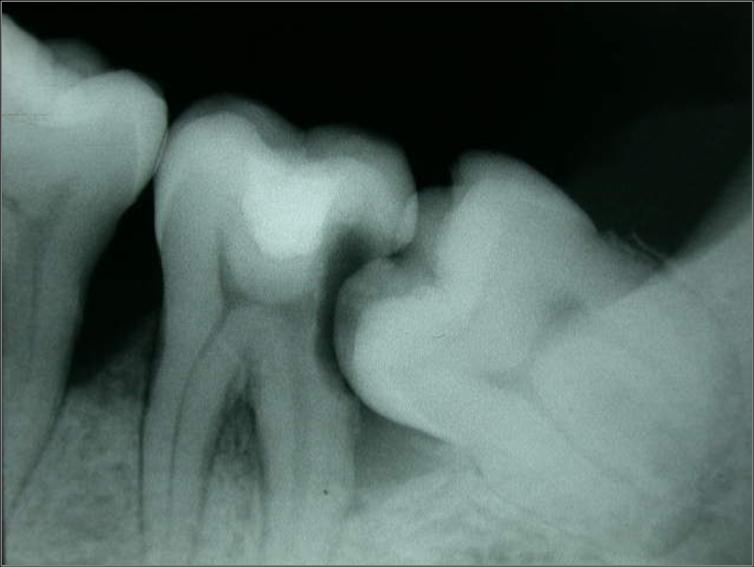
Material and Methods: Five beagle dogs were included in the study. In both quadrants of the mandible, incisions were made in the crevice region of the third and fourth pre-molars. Buccal and minute lingual full-thickness flaps were elevated. The mesial root of the four pre-molars root was filled and the teeth were hemi-sected. Following flap elevation in 3P3 and 4P4 regions, the distal roots were removed. In the right jaw quadrants, implants with a sand blasted and acid etched (SLA) surface were placed in the fresh extraction sockets, while in the left jaws the corresponding sockets were left for spontaneous healing. The mesial roots were retained as surgical control teeth. After 3 months, the animals were examined clinically, sacrificed and tissue blocks containing the implant sites, the adjacent tooth sites (mesial root) and the edentulous socket sites were dissected, prepared for ground sectioning and examined in the microscope.
Results: At implant sites, the level of bone-to-implant contact (BC) was located 2.6±0.4 mm (buccal aspect) and 0.2±0.5 mm (lingual aspect) apical of the SLA level. At the edentulous sites, the mean vertical distance (V) between the marginal termination of the buccal and lingual bone walls was 2.2±0.9 mm. At the surgically treated tooth sites, the mean amount of attachment loss was 0.5±0.5 mm (buccal) and 0.2±0.3 mm (lingual).
Conclusions: Marked dimensional alterations had occurred in the edentulous ridge after 3 months of healing following the extraction of the distal root of mandibular pre-molars. The placement of an implant in the fresh extraction site obviously failed to prevent the re-modelling that occurred in the walls of the socket. The resulting height of the buccal and lingual walls at 3 months was similar at implants and edentulous sites and vertical bone loss was more pronounced at the buccal than at the lingual aspect of the ridge. It is suggested that the resorption of the socket walls that occurs following tooth removal must be considered in conjunction with implant placement in fresh extraction sockets.

Dynamics of bone tissue formation in tooth extraction sites
An experimental study in dogs
G. Cardaropoli1, M. Araújo1,2 and J. Lindhe1
J Clin Periodontol 2003; 30, 809~818

Objectives: The aim of the present experiment was to study events involved in the healing of marginal, central and apical compartments of an extraction socket, from the formation of a blood clot, to bone tissue formation and remodeling of the newly formed hard tissue.
Material and Methods: Nine mongrel dogs were used for the experiment. The fourth mandibular premolars were selected for study and were divided into one mesial and one distal portion. The distal root was removed and the socket with surrounding soft and mineralized tissue was denoted "experimental unit". The dogs were killed 1, 3, 7, 14, 30, 60, 90, 120 and 180 days after the root extractions. Biopsies including the experimental units were demineralized in EDTA, dehydrated in ethanol and embedded in paraffin. Serial sections 7 m thick were cut in a mesio-distal plane. From each biopsy, three sections representing the central part of the socket were selected for histological examination. Morphometric measurements were performed to determine the volume occupied by different types of tissues in the marginal, central and apical compartments of the extraction socket at different intervals.
Results: During the first 3 days of healing, a blood clot was found to occupy most of the extraction site. After seven days this clot was in part replaced with a provisional matrix (PCT). On day 14, the tissue of the socket was comprised of PM and woven bone. On day 30, mineralized bone occupied 88% of the socket volume. This tissue had decreased to 15% on day 180. The portion occupied by bone marrow(BM) in the day 60 specimens was about 75%, but had increased to 85% on day 180.
Conclusion: The healing of an extraction socket involved a series of events including the formation of a coagulum that was replaced by (i) a provisional connective tissue matrix, (ii) woven bone, and (iii) lamellar bone and BM. During the healing process a hard tissue bridge cortical bone formed, which "closed" the socket.

Healing of extraction sockets and surgically produced augmented and non-augmented defects in the alveolar ridge. An experimental study in the dog
G. Cardaropoli1, M. Araújo1,2, R. Hayacibara2, F. Sukekava2 and J. Lindhe1
J Clin Periodontol 2005; 32: 435~440

Objectives: The current experiments had three aims (i) to determine whether the absence of the periodontal ligament (PDL) may alter features of the healing of an extraction socket, (ii) to examine if there were differences in the proportion of different tissues in resolved extraction sockets and surgically produced defects after 3 months of healing, (iii) to study the influence of different biomaterials on the healing of surgically produced bone defects.
Material and Methods: Extraction sites: In five dogs, the 4th mandibular pre-molars were hemi-sected and the distal roots were removed. The extraction socket of one of the pre-molars was instrumented to eliminate all remnants of the PDL tissue. The socket of the contra-lateral pre-molar was left without instrumentation. The dogs were sacrificed after 3 months of healing.
Defect sites: In five dogs, the pre-molars and 1st molars on both sides of the mandible were first removed and 3 months of healing allowed. After this interval three standardized cylindrical defects were prepared in each side of the mandible. The defects were 3.5 mm in diameter and 8 mm deep.
In each quadrant one defect was grafted with Bio-Oss® Collagen, one with Collagen Sponge and one defect was left non-grafted. The dogs were sacrificed 3 months after the grafting procedure.
Results: Extraction sites: The two categories of extraction sockets did not differ with respect to gross morphological features. The tissue of the extraction sites, apical of a newly formed bone bridge, was dominated by bone marrow. Few trabeculae of lamellar bone were also present.
Defect sites: The non-augmented defect was sealed by a hard-tissue bridge. In the central and apical portions of the defect bone marrow made up about 61%, and mineralized bone 39% of the tissues. The invagination of the surface of this crestal bone was 0.8±0.3 mm.
The defect augmented with Collagen Sponge was covered by a hard-tissue bridge 38% of the tissue within the defect was made up of bone marrow while the remaining 62% was occupied by mineralized bone. The invagination of the hard-tissue bridge was on the average 0.6±0.1 mm.
In defects augmented with Bio-Oss® Collagen the biomaterial occupied a substantial portion of the tissue volume. Eighty-five percent of the periphery of the Bio-Oss® particles were found to be in direct contact with newly formed mineralized bone. Woven bone and bone marrow made up 47% and 26% of the newly formed tissue. The invagination of the most coronal part of the bone defect was 0.1±0.1 mm.
Conclusion: Sockets that following tooth removal had their PDL tissue removed exhibited similar features of healing after 3 months as sockets which had the PDL retained. The tissues present in an extraction site appeared to be more mature than those present in a surgically produced defect of similar dimension. The Bio-Oss® Collagen augmented defect exhibited less wound shrinkage than the non-augmented defect.

Dimensional ridge alterations following tooth extraction. An experimental study in the dog
Mauricio G. Araújo1,2 and Jan Lindhe2
J Clin Periodontol 2005; 32: 212~218

Objective: To study dimensional alterations of the alveolar ridge that occurred following tooth extraction as well as processes of bone modelling and remodelling associated with such change.
Material and Methods: Twelve mongrel dogs were included in the study. In both quadrants of the mandible incisions were made in the crevice region of the 3rd and 4th premolars. Minute buccal and lingual full thickness flaps were elevated. The four premolars were hemi-sected. The distal roots were removed. The extraction sites were covered with the mobilized gingival tissue. The extractions of the roots and the sacrifice of the dogs were staggered in such a manner that all dogs contributed with sockets representing 1, 2, 4 and 8 weeks of healing. The animals were sacrificed and tissue blocks containing the extraction socket were dissected, decalcified in EDTA, embedded in paraffin and cut in the buccal lingual plane. The sections were stained in haematoxyline eosine and examined in the microscope.
Results: It was demonstrated that marked dimensional alterations occurred during the first 8 weeks following the extraction of mandibular premolars. Thus, in this interval there was a marked osteoclastic activity resulting in resorption of the crestal region of both the buccal and the lingual bone wall. The reduction of the height of the walls was more pronounced at the buccal than at the lingual aspect of the extraction socket. The height reduction was accompanied by a "horizontal" bone loss that was caused by osteoclasts present in lacunae on the surface of both the buccal and the lingual bone wall.
Conclusions: The resorption of the buccal/lingual walls of the extraction site occurred in two overlapping phases. During phase 1, the bundle bone was resorbed and replaced with woven bone. Since the crest of the buccal bone wall was comprised solely of bundle this modelling resulted in substantial vertical reduction of the buccal crest. Phase 2 included resorption that occurred from the outer surfaces of both bone walls. The reason for this additional bone loss is presently not understood.

Hard-tissue alterations following immediate implant placement in extraction sites
Daniele Botticelli1,2, Tord Berglundh1 and Jan Lindhe1
J Clin Periodontol 2004;32:820~828

Background: The marginal gap that may occur following implant installation in an extraction socket may be resolved by hard-tissue fill during healing.
Objective: To study dimensional alterations of hard tissues that occur following tooth extraction and immediate placement of implants.
Material and methods: Eighteen subjects with a total of 21 teeth scheduled for extraction were included. Following flap elevation and the removal of a tooth and implant installation, clinical measurements were made to characterize the dimension of the surrounding bone walls, as well as the marginal defect. No membranes or filler material was used. The flaps were subsequently replaced and secured with sutures in such a way that the healing cap of the implant was exposed to the oral environment. After 4 months of healing a re-entry procedure was performed and the clinical measurements were repeated.
Results: Fifty-two marginal defects exceeding 3 mm were present at baseline: 21 at buccal, 17 at lingual/palatal, and 14 at approximal surfaces. At the re-entry eight defects exceeding 3.0 mm remained. During the 4 months of healing, the bone walls of the extraction underwent marked change. The horizontal resorption of the buccal bone dimension amounted to about 56%. The corresponding resorption of the lingual/palatal bone was 30%. The vertical bone crest resorption amounted to 0.3±0.6 mm (buccal), 0.6±1.0 mm (lingual/palatal), 0.2±0.7 mm (mesial), and 0.5±0.9 mm (distal).
Conclusion: The marginal gap that occurred between the metal rod and the bone tissue following implant installation in an extraction socket may predictably heal with new bone formation and defect resolution. The current results further documented that marginal gaps in buccal and palatal/lingual locations were resolved through new bone formation from the inside of the defects and substantial bone resorption from the outside of the ridge.

Resolution of bone defects of varying dimension and configuration in the marginal portion of the peri-implant bone
An experimental study in the dog

Daniele Botticelli, Tord Berglundh and Jan Lindhe
J Clin Periodontol 2004; 31: 309~317

Background: It was demonstrated that a marginal defect of about 1 mm between the bone wall and the metal surface after implant installation can heal with a high degree of bone fill and osseointegration.
Objective: The aim of the present animal experiment was to study bone healing at implant sites with hard tissue defects of varying dimensions and configuration.
Material and Methods: Four Labrador dogs were used. All mandibular premolars and first molars were extracted. After 3 months of healing, five experimental sites, two control (C1, C2) and three test (T1, T2, T3) sites, were identified. In all five sites, custom-made implants with a sand-blasted, large-grit, acid-etched (SLA) surface and with an outer dimension of 3.3 10 mm, were used. In site C1, traditional implant installation was performed. In site C2, the marginal 5 mm of the canal, prepared for the implant, was widened to 5.3 mm using a step-drill. Thus, following the installation of the implant, a circumferential gap occurred between the bone tissue and the metal rod that was 5 mm deep and between 1 and 1.25 mm wide. In test site T1, the canal was widened to establish a marginal gap of 2 2.25 mm. In test sites T2 and T3, the marginal 5 mm of the canal was first widened to 5.3 mm (T2) or 7.3 mm (T3). The buccal bone wall opposite the defect was subsequently removed. Following the placement of a cover screw in sites C2, T1, T2, and T3, a resorbable membrane was placed over the defect. All implants were submerged. After 4 months of healing, block biopsies of each implant site were dissected and processed for ground sectioning.
Results: The observations disclosed that four-wall defects of different dimensions (1 2.25 mm wide) that occurred in the marginal portion of the recipient sites following implant installation were resolved during healing. Further, at sites where the buccal bone wall during defect preparation was intentionally removed, healing resulted in defect resolution at the mesial, distal, and lingual aspects. At the buccal aspects, healing was incomplete but the dimension of the defect was reduced by the limited amounts of new bone formation extending from the lateral and apical borders of the defect.
Conclusion: Wide marginal defects may during healing be filled with bone. In such defects a high degree of osseointegration may occur to implants designed with an SLA surface.