Perio-Implant Journals

跟牙周病治療與植牙相關的文獻

My Photo
Name:
Location: 台南市, Taiwan

A tour-crazy periodontist around the World. - I like to drive VOVLE cars and put sweden-made dental implants (Nobelcare & Astra) and live with IKEA furnishes .

Wednesday, January 18, 2006

TGF-Beta Signaling


What are your thoughts on the recent finding, released only last month, that the quality of bone matrix, a key component of bone, is regulated by a molecule known as transforming growth factor beta or TGF-Beta? The research may lead to improvements in the quality and speed of bone repair following dental implant placement or bone grafting.
For those who did not get a chance to read about the study, below is a brief sypnosis:

The ability of bone to resist damage depends on the mass, or quantity, of bone, its architecture and the quality of bone matrix, the mineralized material between cells. Several molecular factors have been shown to regulate the mass and architecture of bone. So far, however, none have been shown to regulate bone matrix, which is responsible for bone elasticity and toughness. There has been significant disagreement about whether the quality of bone matrix varies among individuals and, if it does, whether it could be altered for therapeutic reasons. In any case, until now, scientists have lacked a strategy for measuring its quality and teasing out its impact, says senior study author Tamara Alliston, PhD, UCSF assistant adjunct professor of Cell and Tissue Biology.
In the current study, the team explored whether transforming growth factor beta (TGF-ß) regulates the properties of bone matrix because there were hints that it might. TGF-ß is known to play a role in the development of osteoblasts, cells that produce bone matrix.
The researchers carried out their evaluation in five sets of mice genetically engineered to produce differing levels of TGF-ß signaling within osteoblasts, and, for comparison, in normal, or 'wild type' mice. After the animals had been euthanized, the team utilized highly sensitive instruments developed in the materials sciences -- atomic-force microscopy, x-ray tomography and micro-Raman spectroscopy -- to measure the properties of bone matrix independent of bone mass and architecture. They also compared the bones' resistance to fracture in a bending test.The results were notable, according to Alliston. In animals genetically engineered to produce high levels of TGF-ß, the measurements of bone matrix indicated increased susceptibility to fracture. The matrix was less elastic, less hard and contained lower levels of the mineral calcium phosphate. In addition, the animals' bones were less resistant to fracture in the bending test.In contrast, in animals with low levels of TGF-ß the bone matrix was more elastic, harder, had higher mineral concentration and the bone overall had increased mass. In addition, the bones were more resistant to fracture in the bending test.The bones studied included the femur, tibia and calvarial parietal bones."This is the first evidence that properties of bone matrix can be regulated by a growth factor and that by modifying the TGF-ß pathway, specifically, these properties can be controlled," says Alliston.The study suggests, she says, that TGF-ß could be targeted for clinical intervention in patients. "By decreasing TGF-ß signaling at the relevant site in the body, we may be able to improve the quality of bone to either prevent the damage that occurs in osteoarthritis and osteoporosis, or improve the quality and speed of bone repair following bone fracture, joint implantation, dental implants or bone grafting.""If we could decrease the production of TGF-ß at the site of the transplant, we might be able to strengthen the quality of bone being formed," says the lead author of the study, Guive Balooch, BA, in the UCSF Graduate Program in Oral and Craniofacial Sciences and Division of Bioengineering.

Source: http://pub.ucsf.edu/newsservices/releases/200512136/

Tuesday, January 10, 2006

Localized ridge augmentation using a micro titanium mesh

Localized ridge augmentation using a micro titanium mesh: a report on 27 implants followed from 1 to 3 years after functional loading.

von Arx T, Wallkamm B, Hardt N.
Department of Oral and Maxillofacial Surgery, Kantonsspital, Lucerne, Switzerland.

Clin Oral Implants Res. 1998 Apr;9(2):123-30

The present paper describes the clinical and radiographic healing results of 27 implants followed from 1 to 3 years after functional implant loading. Prior to implant placement, alveolar ridges with insufficient bone volume were augmented using autogenous bone grafts and a micro titanium mesh for graft stabilization. After a mean interval of 5.2 months implants were installed. Following an osseointegration period of on average 7.2 months, implants were supplied with suprastructures. The mean loading period for the 27 implants was 21 months. All implants exhibited ankylotic stability and healthy peri-implant soft tissues. The detailed analysis of clinical parameters (probing depth, level of mucosal margin, attachment level, modified plaque and sulcus bleeding indices) and radiographic measurements (crestal bone level), revealed findings similar to those at implants placed into non-augmented bone. Peri-implant bone resorption was calculated to be 1.0 mm for the 1st year after implant loading and 0.1 mm for the following year. Pain, suppuration or semilunar bone defects were absent at all implants. It was concluded that loaded dental implants which have been inserted into an augmented alveolar ridge using autogenous bone grafts and a micro titanium mesh for graft stabilization, demonstrate clinical and radiographic findings similar to those of implants placed into a pristine ridge.

Treatment of periimplantitis with an enamel matrix protein derivative

T.M. AUSCHILL1, P. WINDISCH2, F. DÖRI2, and A. SCULEAN3,
1 Albert-Ludwigs-University Freiburg, Germany, 2 Semmelweis University, Budapest, Hungary, 3 University of Saarland, Homburg, Germany

The topical application of an enamel matrix protein derivative (EMD) onto periodontally compromised root surfaces has been shown to promote periodontal wound healing and regeneration. However, up to now no data are available on the healing of periimplantitis defects following treatment with EMD.

Objectives: To evaluate the effectiveness of EMD in the surgical treatment of periimplantitis.

Methods: A total of eight patients with one periimplantitis defect each were enrolled into the study. Following the elevation of full thickness mucoperiosteal flaps and careful removal of granulation tissue the implant surfaces were conditioned for 3 min. with a 24% EDTA gel. After rinsing with sterile saline in order to remove the EDTA residues EMD (Emdogain®, BIORA AB, Malmö, Sweden) was applied onto the implant surfaces and into the defects.

Results: At one year after surgery mean probing depth (PD) was reduced from 7.1 ± 1.5 mm to 3.5 ± 1.6 mm (p<0.001)>Conclusion: The present preliminary data suggest that the use of EMD after surgical treatment of periimplantitis leads to significant PD reduction, CAL gain and radiographic bone fill. Controlled histological and clinical studies are needed in order to further elucidate the relevance of this therapy for the treatment of periimplantitis.

Wednesday, January 04, 2006

Third molars may have a negative impact on periodontal health


Evidence-Based Dentistry (2005) 6, 95.

Graham J Smart

Design This was a cross-sectional study.
Materials and methods
Data were within a cohort study obtained between 1996 and 1999 on 6793 people of 52–74 years of age from the dental substudy of the Atherosclerosis Risk in Communities Study. The main independent variable was presence or absence of third molars, assessed visually, and the dependent variable was assessment of periodontal disease designated by pocket depth of 5 mm or greater (PD5+). Associations were determined using odds ratios and 95% confidence intervals (CI). Weighted multivariable models were fitted using logistic regression and variances were adjusted to account for the clustering of quadrants within individual subjects.
Results
A visible third molar was associated with 1.5-fold (95% CI, 1.4–1.7) the odds of PD5+ on the adjacent second molar, while controlling for other factors associated with the presence of third molars and periodontal disease. Other factors positively associated with PD5+ in the model were male gender, older age, smoking, and irregular and episodic dental visits.
Conclusions
The finding of more severe periodontal conditions associated with visible third molars in these middle-aged and older adults indicates that third molars may continue to have a negative impact on periodontal health well into later life. The relationship between third molars and periodontal disease pathogenesis deserves further study using longitudinal data.

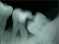
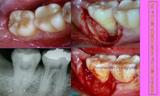
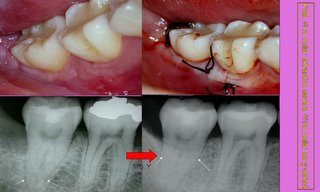
Severe infrabony defect caused from impacted 3rd molar>>Regeneration with Emdogain + Dynagraft. Case report by Perio@